English
Electrocardiography (ECG) is a methodology for recording electrical impulses in the heart. Electrodes measure the depolarisation of the heart and its direction. This can be used to gain a lot of information regarding the distribution of electrical impulses and the problems that may be found there, such as an acute myocardial infarction (current or old), if a patient has ischaemic heart disease or the heart is beating in an irregular rhythm (arrhythmia).
We are not going into detail about how this all works, but will briefly explain the main characteristics of the ECG.
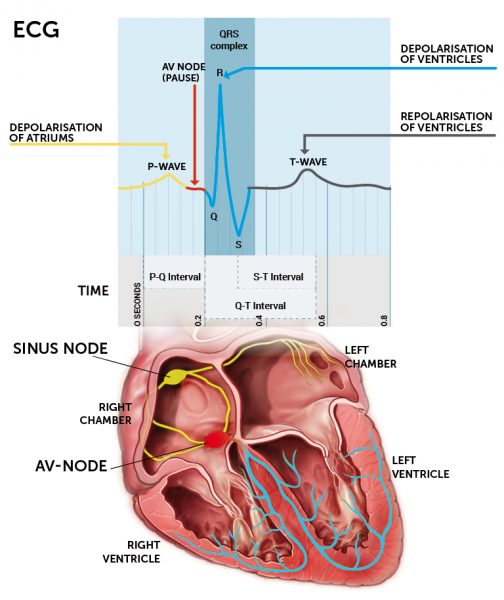
A normal ECG has three different stages. It has a P-wave, a QRS-complex and it a T-wave. Based on this, we get three intervals:
- a P-R interval: the time between the P and R waves
- an S-T interval: the time between the S and T waves
- a Q-T interval: the time from the Q to T waves
The P-wave describes the electrical impulse that runs through the atria. The QRS-complex describes the electrical impulses that run through the ventricles, whereas the T-wave describes the repolarisation of the ventricles, i.e. when the muscle cells of the ventricle recover to its resting potential. Repolarisation of the atria is not shown in the ECG because it occurs while the ventricles are depolarised. The electrical impulses in the ventricles are so much stronger that they ”drown” the impact of the repolarisation of the atria.
Using this knowledge of an ECG, we can look at some simple examples of what can be read from such a recording.
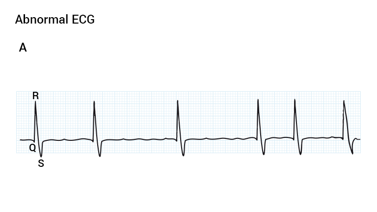
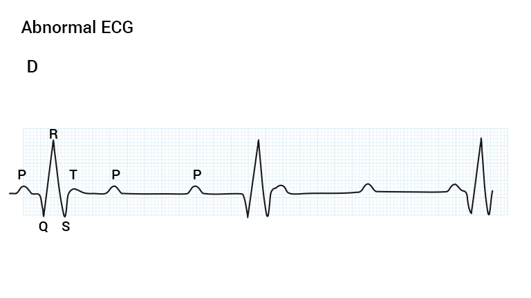
If the atria does not have a coordinated impulse, the muscle cells of the atria all contract independently of each other causing fibrillation. On an ECG there will be no discernible p-waves and an irregular rhythm due to the random stimulation of the AV node.
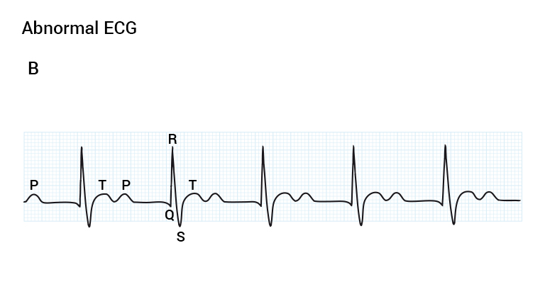
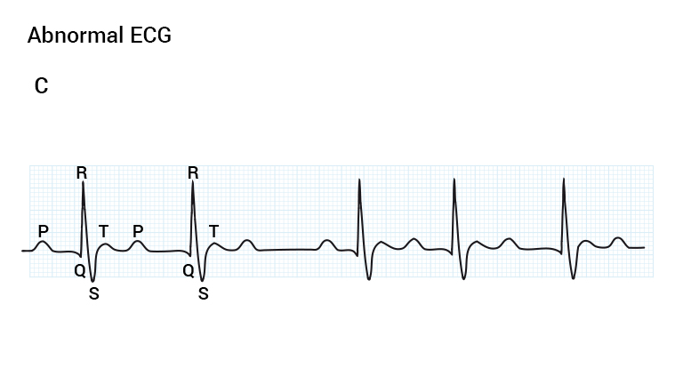
If the PR interval increases, it indicates that the electrical impulses from the atria to the bundle of His are abnormal and there is a delay of transmission to the ventricles, i.e. a prolonged delay in the AV node. This is called an incomplete heart block, and inhibits the heart’s normal activity. In other cases, the P waves and QRS complexes may be completely independent of each other. This indicates complete heart block, where none of the atrial impulses are transmitted through the AV node to the ventricles. In such cases, it is the pacemaker cells of the ventricles that maintain the pumping of blood.
Atrial fibrillation is a condition in which the depolarisation of the atria does not follow the normal pattern, but is completely out of control. The atria do not contract synchronously, but rather vibrate without any particular pump power. On an ECG P waves are absent, and the QRS-complex (ventricular contraction) occurs irregularly because the signal transmission to the ventricles does not follow the normal pattern. The AV-node will thus receive electrical impulses from the atria randomly because of unsynchronized depolarisations of the atria.