English
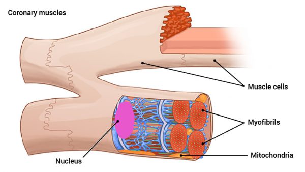
The muscle cells in the heart share several similarities with muscle fibres in skeletal muscles. Note that in skeletal muscles, the long muscle cells are called muscle fibres, while in the heart they’re called cells. As with skeletal muscle fibres, the heart muscle cells have sarcomeres, myofilaments and the sliding actin- and myosin filaments. Actin and Myosin are the proteins which are responsible for the contraction of muscle when myosin filaments slide long actin filaments, secondary to increases in calcium (Ca2+) and ATP concentrations.
In contrast to skeletal muscles, cardiac cells are not elongated. The short cardiac muscle cells are branched. The cardiac contraction follows a pattern where it begins with the atria and spreads through the ventricles, in a specific pattern through the hollow muscle organ. This is achieved with the branched muscle cells.
It is also important to note that the various short muscle cells are connected to each other. They communicate with each other through the cell membranes’ pores (gap junctions), that transmit electrical current between neighbouring cells. The depolarisation leads to muscle contraction starting at one end of the myocardium, the electrical current and contractions spread from muscle cell to muscle cell throughout the heart. This makes the heart contract as a coordinated unit.