English
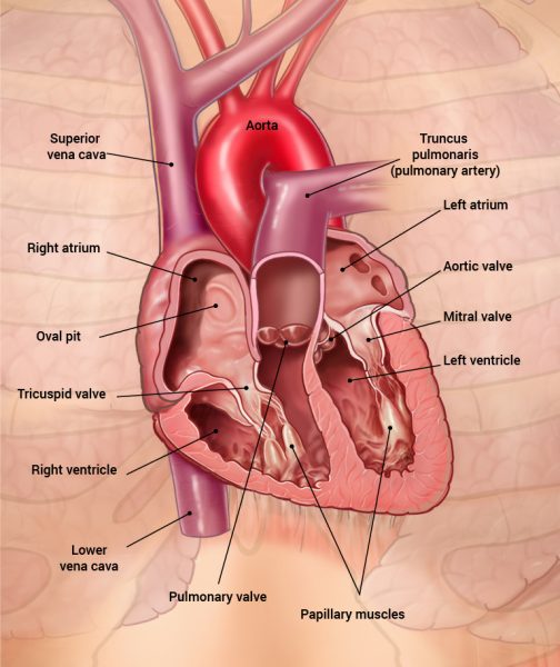
The heart has four heart valves:
- The tricuspid valve lies between the right atrium and right ventricle
- The mitral valve (Bicuspid valve) is located between the left atrium and left ventricle
- The pulmonary valve is located at the transition between the right ventricle and it’s connecting artery, the pulmonary trunk.
- The aortic valve is located at the transition between the left ventricle and the aorta.
Heart valves prevent blood from flowing in the wrong direction when the heart is contracting. As the ventricles contract from the apex upwards (known as systole) and the pressure inside them increases, the valves between the atria and ventricles (atrioventricular valves) are forced shut, preventing backflow of blood (known as regurgitation) into the atria. Simultaneously the pulmonary and aortic valves between the ventricles and blood vessels open. The blood is pushed from the heart and into the pulmonary and systemic circulation.
When the heart relaxes (known as diastole) negative pressure in the ventricles draws blood back from the circulation filling the atria. The negative pressure forces the closure of the aortic and pulmonary valves, preventing regurgitation of blood into the ventricles
The closing of the valves happens quite quickly and it occurs at a high pressure. When we hear the heartbeat, we hear the sound of the valves shutting. We know that when we listen to the heart, we hear two sounds one after another (lub-dub), then a short pause, and then again two sounds. The first of the two sounds you hear is when the valves between the atria and ventricles close, and the other sound comes when the valves between the ventricles and blood vessels close.
The ventricles contain small papillary muscles that attach to the atrioventricular valves. They prevent the valves inverting during systole.